

What is Biomimetic?
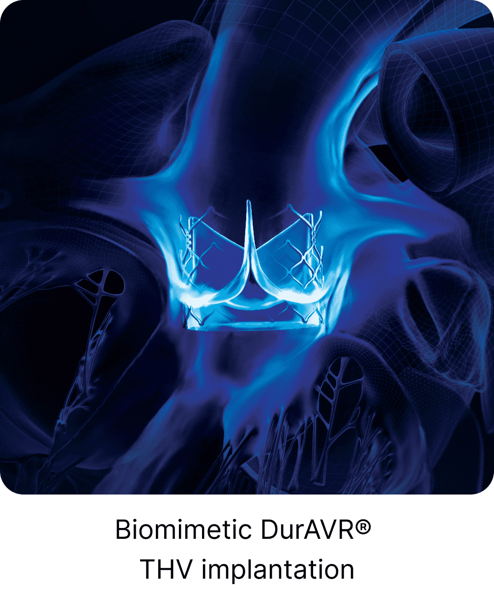
Anteris Chief Medical Officer Christopher Meduri explains the rationale behind the unique single-piece, native-shaped design of the DurAVR® Transcatheter Heart Valve.

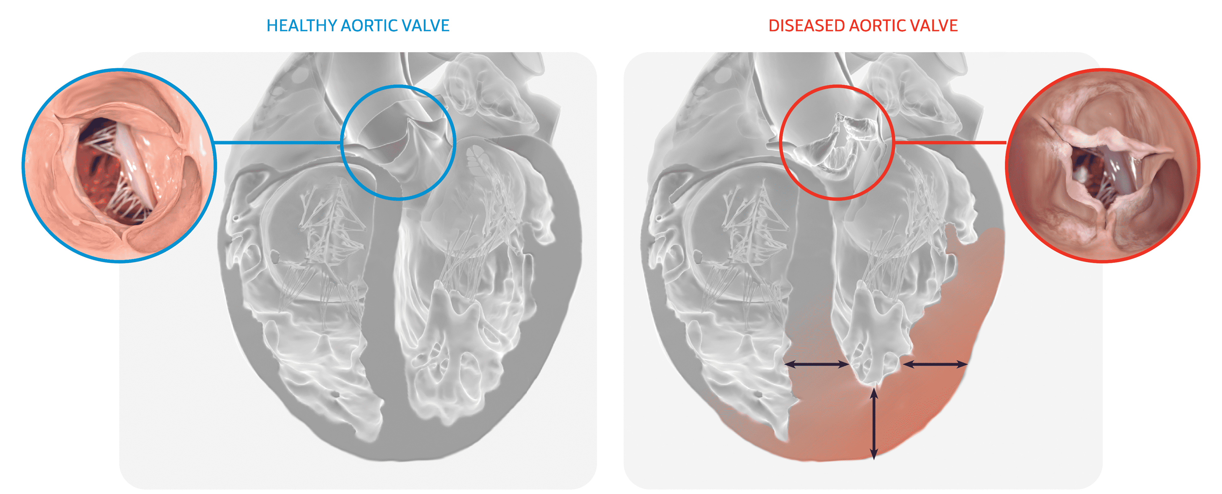
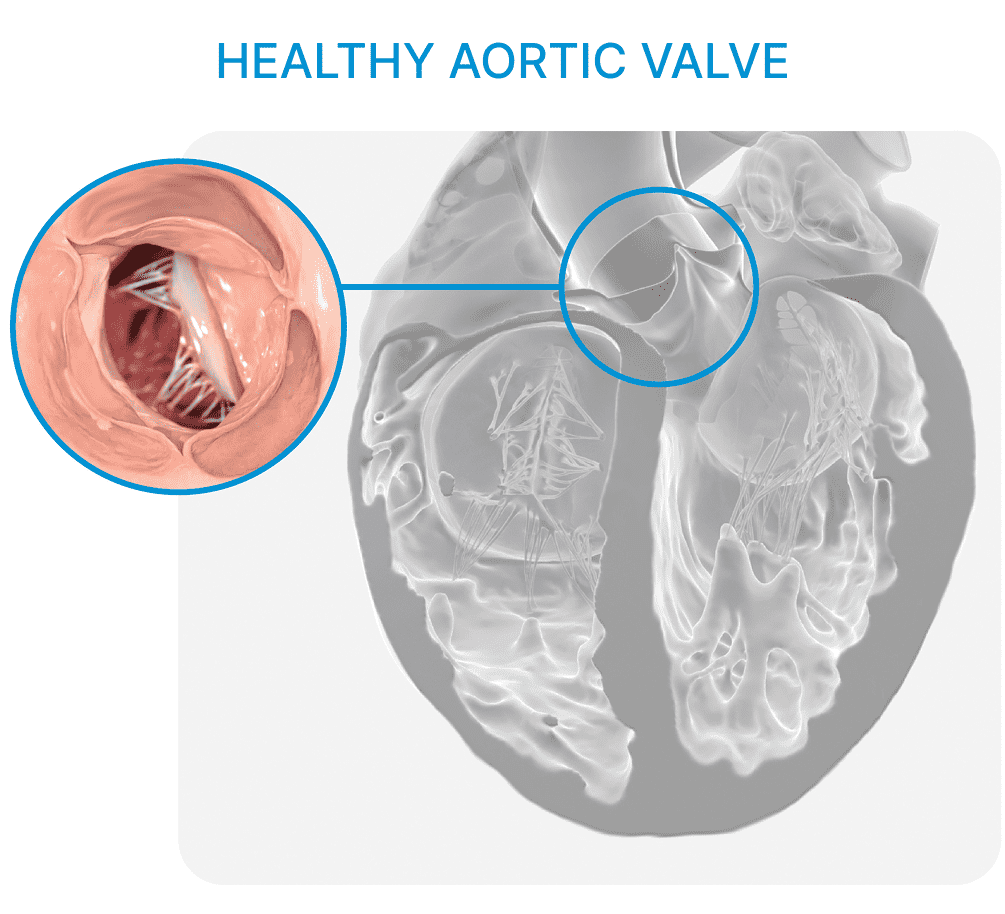
Aortic stenosis (AS) extends beyond being solely a valvular condition; it also affects other parts of the cardiovascular system, influencing both upstream and downstream structures such as the arteries and the heart muscle.
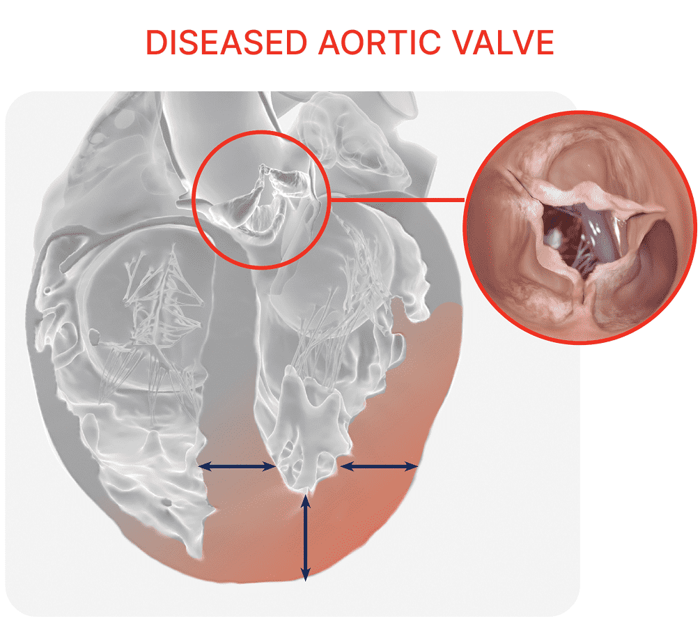
LV Hypertrophy
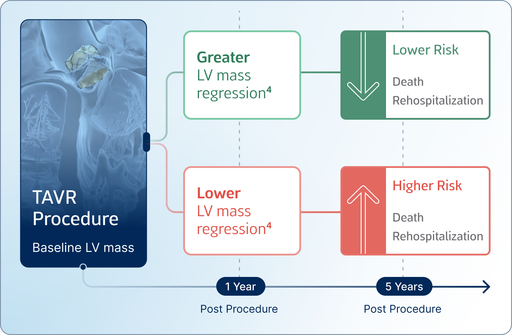
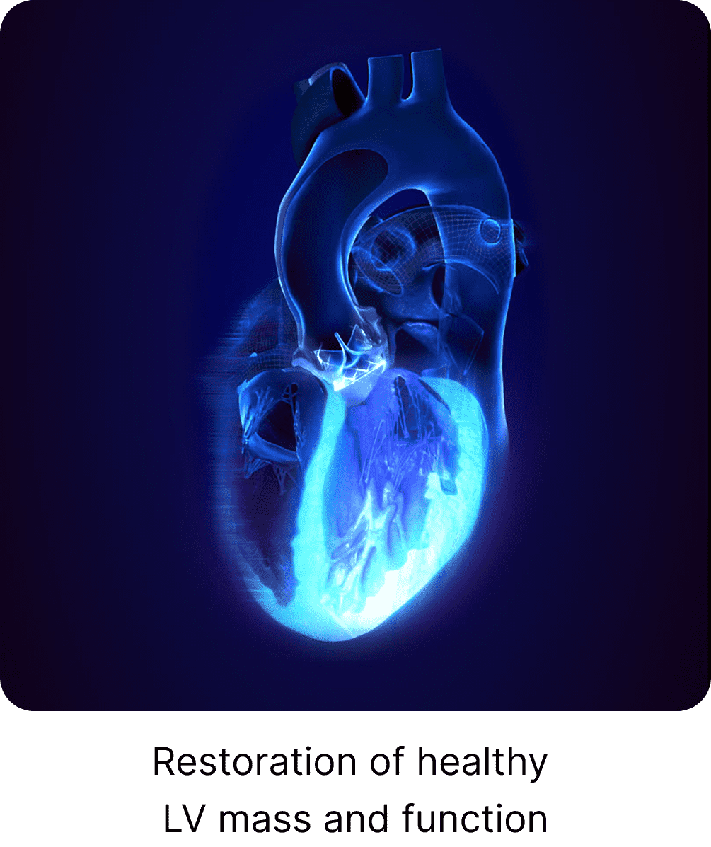
The impact of left ventricular hypertrophy on patient outcomes has already been demonstrated in analyses from the PARTNER trials. Greater left ventricular (LV) mass before TAVR is linked to increased mortality and rehospitalization at five years.3 In addition, a greater reduction in LV mass one year after TAVR is associated with improved long-term outcomes.4
Mean pressure gradients (MPG) are helpful and easy measurements before and after a TAVR procedure. However, they don't tell the full story when it comes to optimal flow. Patients may have good gradients (<10 mmHg) following a TAVR procedure, but they may still experience symptoms and may not experience LV mass regression. Flow dynamics may be an important missing piece of the puzzle.
Laminar Flow Associated with Improved LV Mass Regression
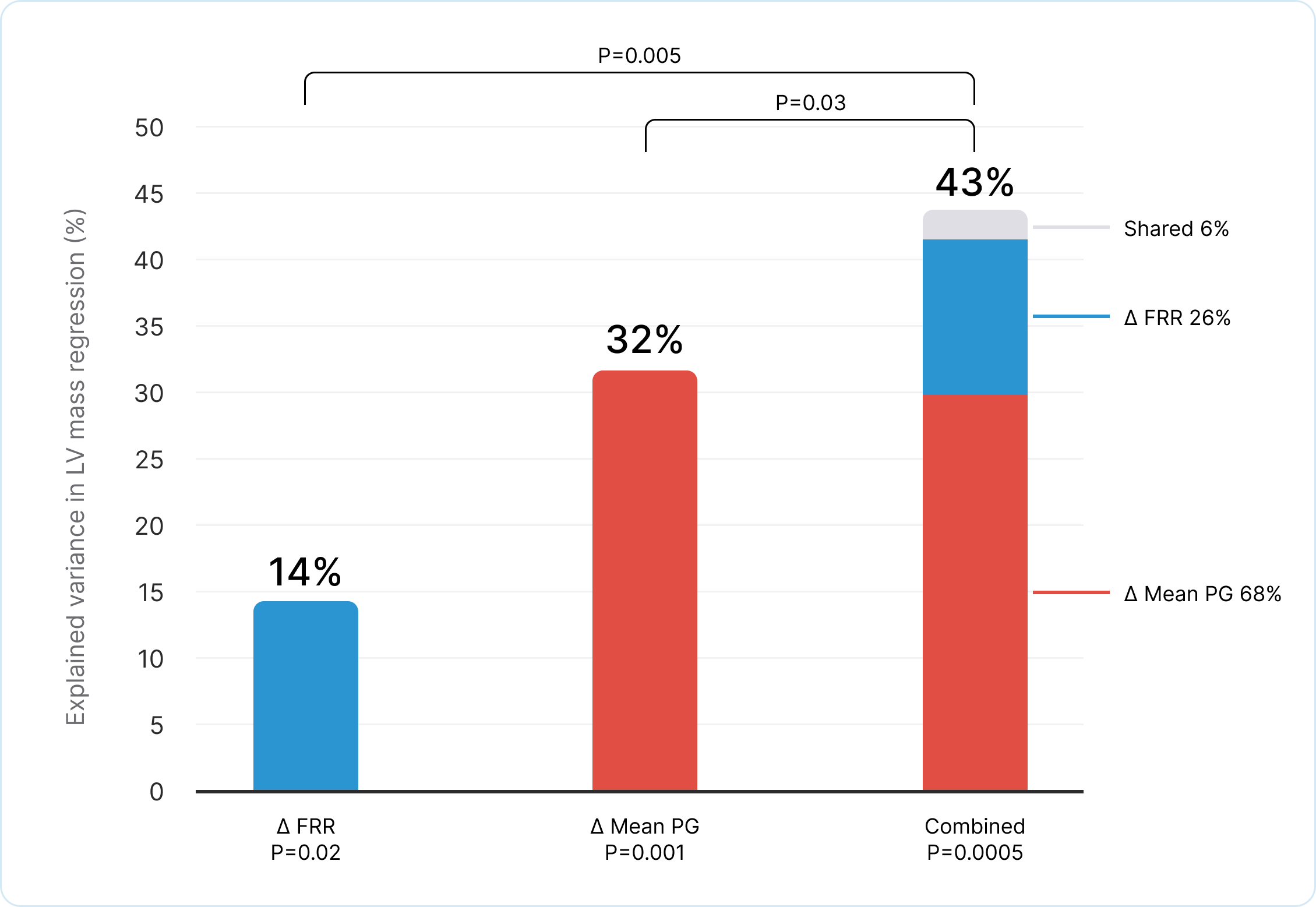
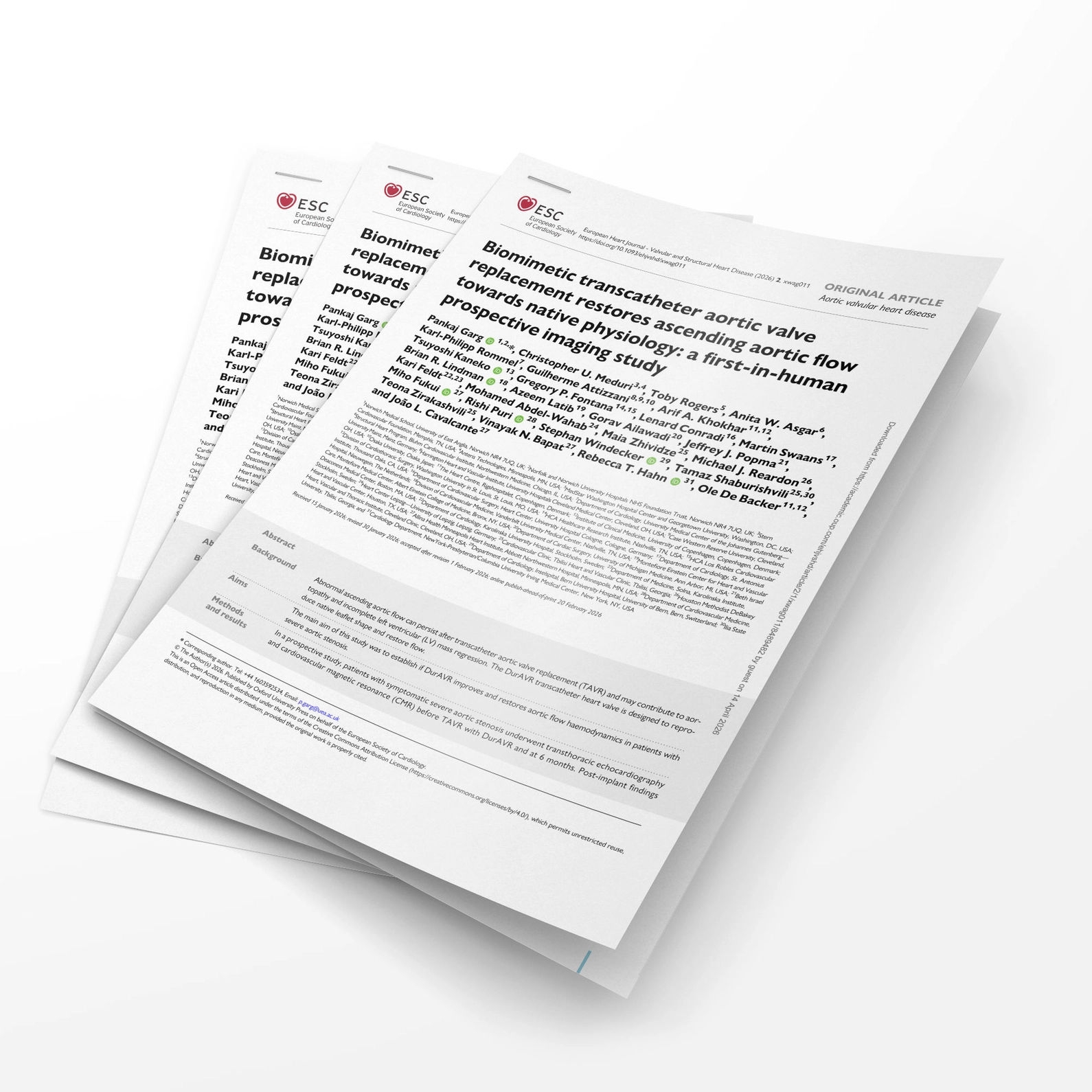
In a statistical analysis of echo and CMR data from 39 paired patients pre-and post-DurAVR® implantation, both change in mean pressure gradient AND change in flow reversal ratio were significantly and independently correlated with the extent of LV mass regression. (Endpoint: ≥10g LV mass regression)5
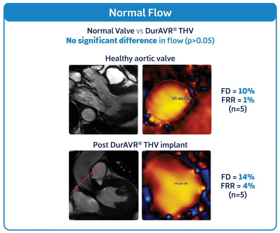
When compared to a healthy aortic valve using cardiac MRI, DurAVR® THV showed no significant difference in flow.
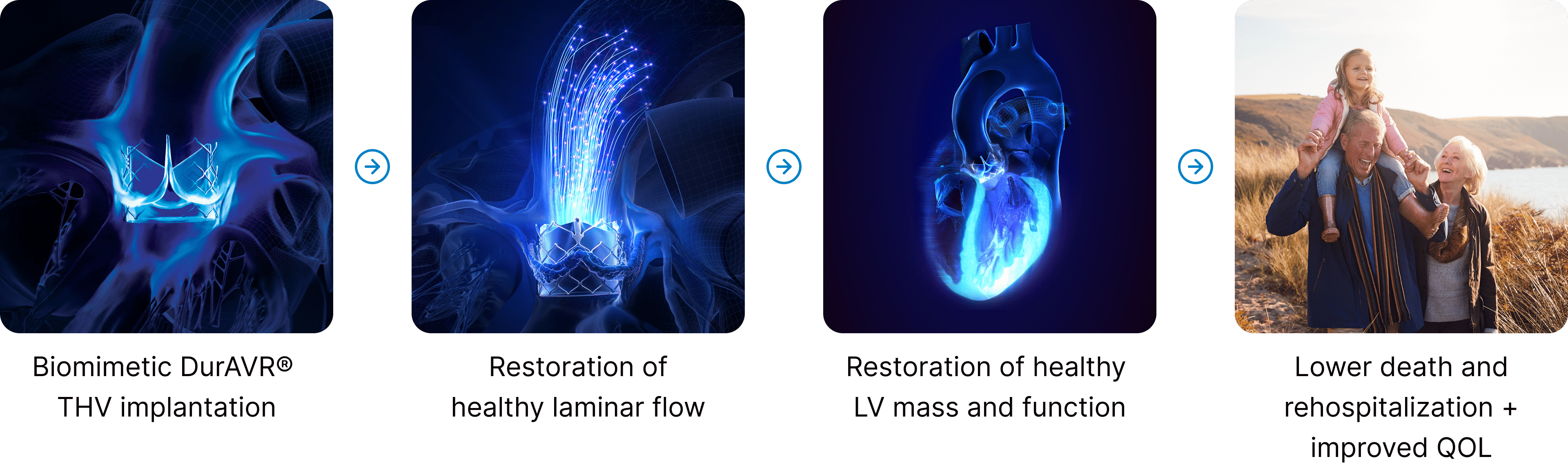
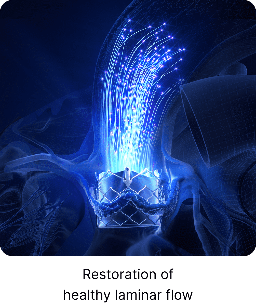
In addition, baseline 4D cardiac MRI from the DurAVR® THV EFS study shows turbulent flow with helical and vortical flow patterns, while imagery taken 6 months post-DurAVR® THV implantation shows evidence of healthy laminar flow through the valve and into the ascending aorta. Cardiac MRI is considered the gold standard for assessing and quantifying valve flow dynamics.
DurAVR® THV has demonstrated a restorative effect on Left Ventricular Mass
DurAVR® THV has demonstrated a restorative effect on Left Ventricular Mass
Patients treated with DurAVR® THV have experienced significant left ventricular (LV) mass regression, measured on cardiac MRI. Matched analysis also confirmed that the LV mass after treatment with DurAVR® THV was comparable to levels seen in healthy controls.
Patients treated with DurAVR® THV have experienced significant left ventricular (LV) mass regression, measured on cardiac MRI. Matched analysis also confirmed that the LV mass after treatment with DurAVR® THV was comparable to levels seen in healthy controls.
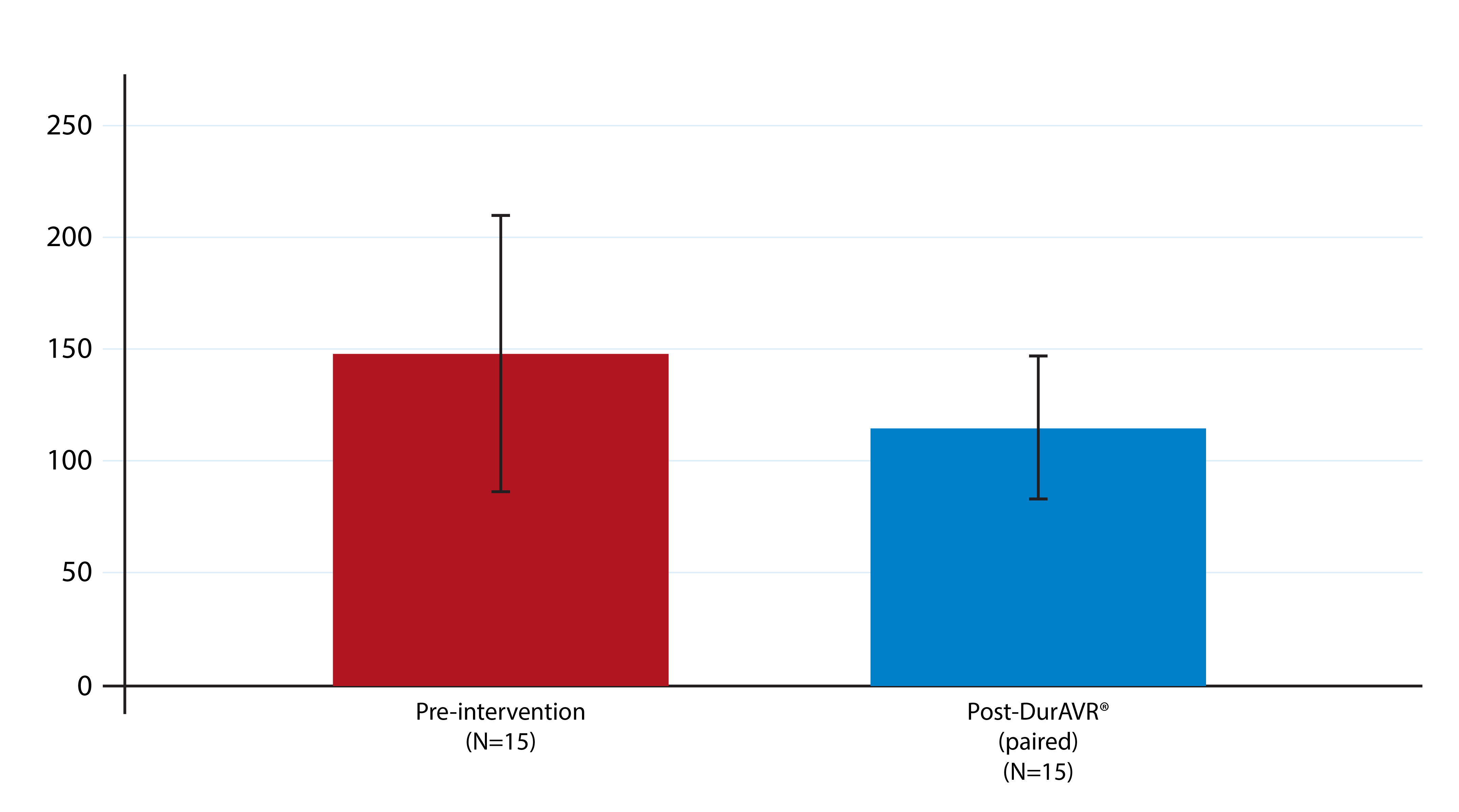
LV Mass
(g)
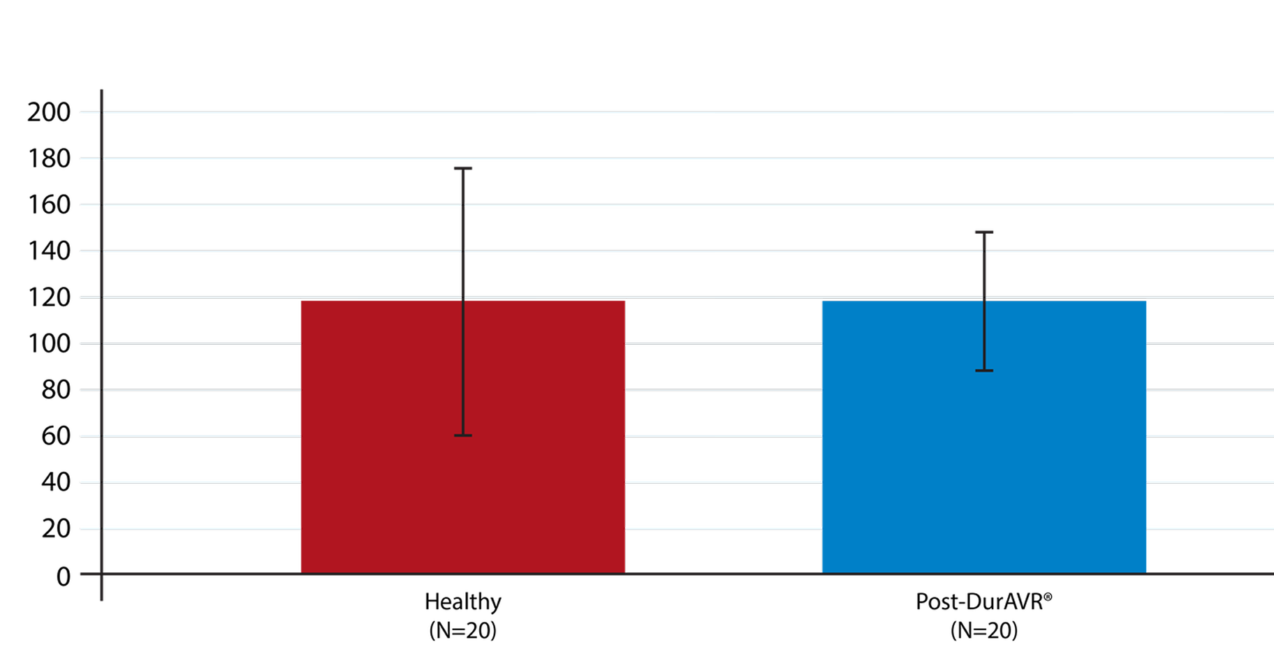
LV Mass
(g)
FIH Study, Presented at TCT 2024


Check out these resources to dive deeper into key topics like laminar flow and LV mass regression.
Watch these short video clips with imaging expert João Cavalcante and Anteris Chief Medical Officer Christopher Meduri.
Anteris Chief Medical Officer Christopher Meduri explains the rationale behind the unique single-piece, native-shaped design of the DurAVR® Transcatheter Heart Valve.

João Cavalcante explains how patients may feel after reverse LV remodeling following a valve replacement.

João Cavalcante summarizes the physiologic mechanisms that contribute to LV hypertrophy.

João Cavalcante explains how aortic stenosis has both upstream and downstream impacts apart from the valve.
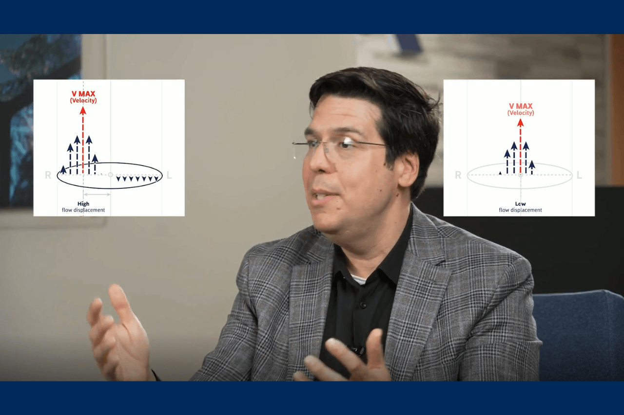
João Cavalcante describes the two key cardiac MRI metrics imagers use to quantify aortic flow patterns.

Imaging expert João Cavalcante explains flow dynamics and what flow patterns are seen in healthy vs. stenotic valves.